
Welcome to Premier Dental of Gettysburg
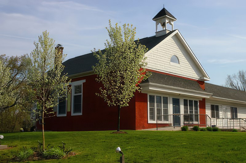
Welcome to Premier Dentistry of Gettysburg, where exceptional dental care meets unparalleled comfort and service. Nestled in the historic Gettysburg area, our practice is dedicated to providing comprehensive dental solutions tailored to the unique needs of each patient.
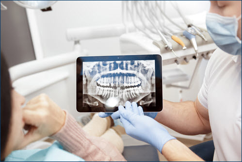
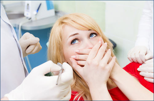
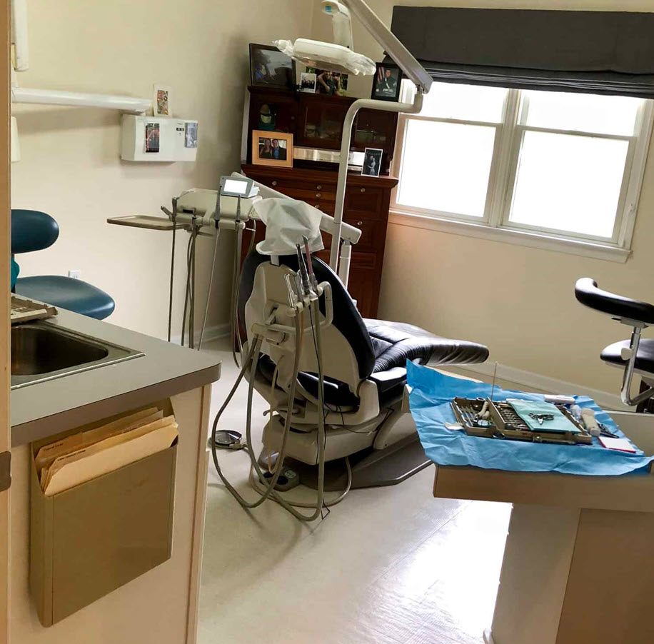
At Premier Dentistry of Gettysburg, we believe in a patient-centered approach. Our state-of-the-art facility is equipped with the latest dental technology, ensuring every procedure from routine check-ups to advanced treatments is performed with utmost precision and care. Our team of experienced dentists and caring staff are committed to making your visit as comfortable and anxiety-free as possible.
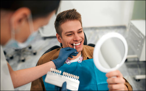
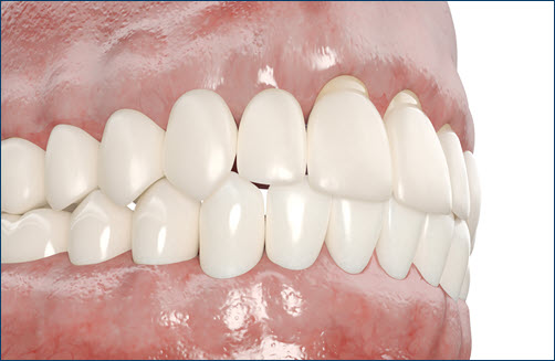
Our services encompass a wide range of dental care, including preventive, cosmetic, restorative, and emergency dentistry. Whether you’re seeking a stunning smile makeover, need a complex dental procedure, or simply require a routine cleaning, we’re here to provide top-notch care. We pride ourselves on our ability to offer advanced treatments like dental implants, Invisalign, and teeth whitening, all under one roof.
Understanding the importance of oral health in your overall well-being, we take the time to educate our patients about dental care and hygiene. Our goal is not just to treat dental issues but to partner with you in maintaining a healthy, beautiful smile for life.
We welcome patients of all ages, offering a warm, family-friendly environment. Your smile is our priority. We invite you to experience the difference in dental care where quality, comfort, and patient satisfaction are at the core of what we do.
Book your appointment today and take the first step towards a healthier, happier smile!
CONTACT INFORMATION:
Address:
1275 Biglerville Road
Gettysburg, PA 17325
Phone: 717-338-9905
Fax: 717-338-9906
Email: Office@PDGettysburg.com
DENTAL CLINIC HOURS:
Monday: 7:30am-5:00pm
Tuesday: 7:30am-5:00pm
Wednesday: 7:30am-5:00pm
Thursday: 7:30am-5:00pm
Friday: Closed
BLOG ARTICLES OF INTEREST: